Biliary hamartoma
Biliary hamartomas (also known as von Meyenburg complexes) are benign malformations of the biliary tract that appear as small, cystic lesions scattered throughout the liver parenchyma. These developmental anomalies result from failure of remodeling of the ductal plates during embryogenesis.
Clinical presentation
Due to their small sizes, Biliary hamartomas are asymptomatic and usually found incidentally. However, in rare cases, they can potentially cause abdominal pain, jaundice, fever, or portal hypertension if they become large enough to obstruct bile flow or significantly impact liver function
Ultrasound Features
- Multiple small lesions (usually 0.1-1.5 cm in diameter)
- Uniform appearance throughout the liver
- Two morphological patterns:
- Cystic pattern: Anechoic with posterior acoustic enhancement
- Solid-appearing pattern: Hyperechoic or hypoechoic nodules
- No vascular flow on Doppler examination
- No mass effect on surrounding vessels
- Diffuse distribution (though may occasionally cluster in one lobe)
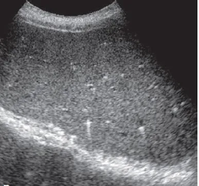
Typical ultrasound appearance of multiple small biliary hamartomas
Advanced Ultrasound Techniques
- Contrast-Enhanced Ultrasound (CEUS)
- No enhancement in cystic lesions
- Possible thin peripheral enhancement in solid-appearing variants
- No late washout (helps differentiate from metastases)
- Elastography
- Normal liver stiffness measurements
- Lesions themselves are too small to assess individually
Clinical Significance
- Entirely benign with no malignant potential
- Usually asymptomatic (incidental finding)
- No surveillance or follow-up needed when typical features present
- Rare associations:
- utosomal dominant polycystic kidney disease
- Congenital hepatic fibrosis
References
- Mortelé KJ, Ros PR. Cystic focal liver lesions in the adult: differential CT and MR imaging features. Radiographics. 2001;21(4):895-910.
- Horton KM, Bluemke DA, Hruban RH, et al. CT and MR imaging of benign hepatic and biliary tumors. Radiographics. 1999;19(2):431-51.
- Choi BI, Yeon KM, Kim SH, Han MC. Caroli disease: central dot sign in CT. Radiology. 1990;174(1):161-3.
- Dietrich CF, Mertens JC, Braden B, et al. Contrast-enhanced ultrasound of histologically proven liver hemangiomas. Hepatology. 2007;45(5):1139-45.