Fetal Sex Determination: Ultrasound Markers and Diagnostic Accuracy
For many expecting parents, discovering the biological sex of their baby is one of the most anticipated milestones of pregnancy. While modern non-invasive prenatal testing (NIPT) can identify fetal sex via cell-free maternal DNA as early as week 10, obstetric ultrasound remains the most widely accessible, real-time method for fetal sex evaluation.
However, the accuracy of ultrasound depends heavily on gestational age, fetal positioning, and the specific anatomical markers being observed.
The First Trimester: Heartbeat Myths vs. Early Anatomical Markers
The first trimester (up to 13 weeks and 6 days) is a period of rapid embryonic development. While parents are often eager for answers during early scans, true anatomical differentiation happens later than most realize.
The Fetal Heartbeat Myth
A widespread piece of pregnancy folklore suggests that fetal heart rate (FHR) can predict a baby’s sex:
- The Claim: A heart rate above 140–150 beats per minute (bpm) indicates a girl, while a lower heart rate indicates a boy.
- The Reality: There is no scientific correlation between first-trimester fetal heart rate and biological sex. During the first trimester, the fetal heart rate starts around 90–110 bpm at week 6, accelerates rapidly to a peak of 170–180 bpm by week 9, and then gradually plateaus between 120–160 bpm by the end of the first trimester. Multiple clinical studies have analyzed thousands of scans and found no statistically significant difference between male and female heart rates during early pregnancy. FHR reflects gestational age and cardiac development, not biological sex.
True First-Trimester Determinants: The Sagittal Sign (Nub Theory)
While genital organs look virtually identical externally until around week 9, an early anatomical feature called the genital tubercle (or “nub”) forms in both sexes.
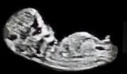
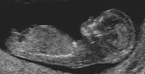
By weeks 11 to 13, sonographers can use a high-resolution midsagittal plane view to assess the angle of this tubercle relative to the fetal spine—a method known clinically as the Sagittal Sign or colloquially as the “Nub Theory”.
- Male Fetus: The genital tubercle typically points cranially (upward), forming an angle of greater than 30 degrees relative to a line drawn parallel to the lower spine.
- Female Fetus: The tubercle points caudally (downward or horizontally), parallel to the spine, at an angle of less than 10 degrees (or less than 30 degrees with no upward tilt).
The Second Trimester: The Gold Standard for Sex Determination
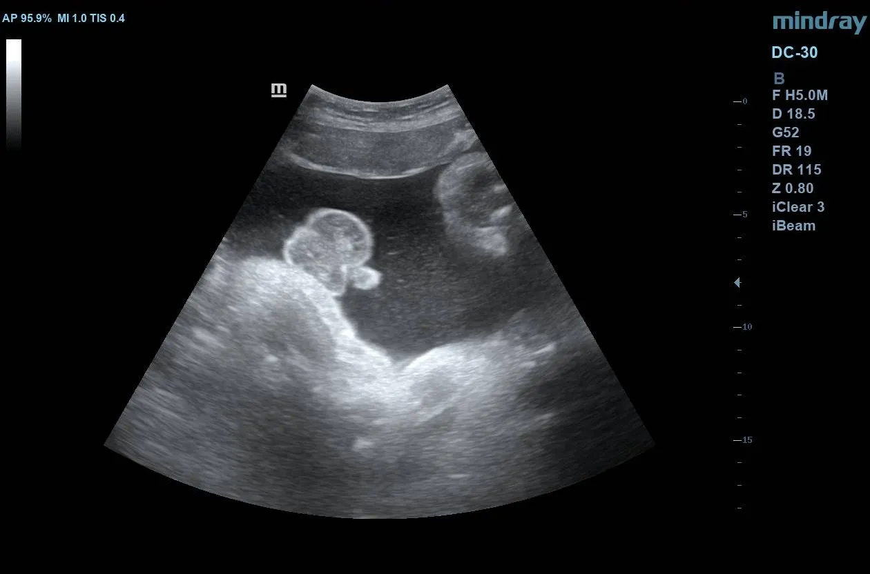
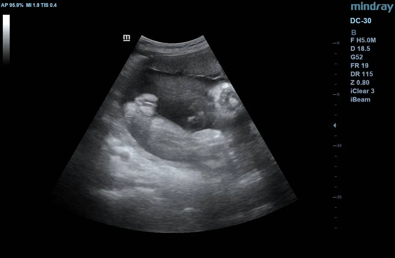
The second trimester—specifically during the routine mid-trimester anomaly scan (18 to 22 weeks)—is the most reliable window for ultrasound sex determination. By this stage, external genitalia are fully differentiated and clearly visible in favorable fetal positions.
Sonographers look for specific, distinct structural signs using a transverse perineal view (often referred to as the pelvic or “potty shot”):
- The Male Determinant (The “Turtle Sign”): In the second trimester, the presence of the penis and scrotum yields a distinct profile. When imaging from below the fetal pelvis, the penis and surrounding scrotal sac resemble the silhouette of a turtle’s head emerging from its shell.
- The Female Determinant (The “Hamburger Sign”): Conversely, the absence of male genitalia and the presence of female structures create a parallel presentation. The sonographer identifies the labia majora and minora, which appear as three distinct white parallel lines on the ultrasound screen, frequently referred to as the “hamburger sign.”
:::note[Key Diagnostic Points]
- Fetal heart rate is entirely inaccurate for sex determination and should never be used clinically.
- The mid-trimester anomaly scan (18–22 weeks) remains the diagnostic gold standard for ultrasound-based sex determination, yielding nearly 100% accuracy.
- Persistent fetal positioning, low amniotic fluid, or high maternal BMI are the primary limitations to capturing an optimal view. :::
The Third Trimester: Confirmation and Structural Validation
By the third trimester (28 weeks onward), sex determination is rarely the primary objective of an obstetric scan, which focuses instead on fetal growth, amniotic fluid volume, and placental positioning. However, confirming or reassessing sex during this period is highly straightforward due to advanced fetal maturity.
Third-Trimester Structural Markers
- Male Fetuses: The sonographer can often visualize the descent of the testes into the scrotal sac, a process that typically begins around week 26 to 28 and progresses throughout the third trimester. Fluid within the scrotum (transient hydrocele) is also a common, normal finding that makes identification very clear.
- Female Fetuses: The labia and clitoris remain distinct, though late-pregnancy crowding can sometimes make capturing a clear perineal view more physically challenging than in the second trimester.
Challenges of Late-Pregnancy Imaging
While the anatomy is fully formed, the sheer size of the fetus relative to the uterine cavity in the third trimester means there is less amniotic fluid space to manipulate the transducer for a clear view. If the baby is deeply engaged in the pelvis or in a persistent breech position, visualizing the perineum can be significantly more difficult than it was at 20 weeks.
Summary of Ultrasound Sex Determinants
| Gestational Window | Primary Ultrasound Marker / Sign | Clinical Accuracy | Diagnostic Limitations |
|---|---|---|---|
| First Trimester (11–13 Weeks) | Sagittal Sign / Genital Tubercle Angle (Nub Theory). | Moderate to High (70%–95%) | Highly dependent on gestational age and neutral fetal positioning. |
| Second Trimester (18–22 Weeks) | “Turtle Sign” (Male) vs. “Hamburger Sign” (Female) via transverse view. | Very High (~99%) | Fetal position (legs crossed, breech), cord location, high maternal BMI. |
| Third Trimester (28+ Weeks) | Testicular descent into scrotum (Male) vs. mature labial structures (Female). | Excellent | Restricted view due to late-stage fetal crowding and pelvic engagement. |