Glycogen Storage Disease (GSD): Ultrasound Diagnosis & Management
Glycogen storage diseases (GSDs) are a group of inherited metabolic disorders characterized by abnormal glycogen metabolism. Hepatic involvement is prominent in types I (von Gierke disease), III (Cori disease), IV, VI, and IX. Ultrasound plays a key role in the initial evaluation and long-term monitoring of hepatic complications.
Ultrasound Features
Characteristic sonographic findings seen across hepatic glycogen storage diseases include:
- Marked Hepatomegaly: Liver span length reads greater than 15 cm in the midclavicular line. It presents as a diffuse, homogeneous enlargement during early stages, and is most prominent in GSD types I and III.
- Increased Parenchymal Echogenicity: Demonstrates a distinct “bright liver” appearance. This occurs due to progressive intracellular glycogen accumulation. It is important to differentiate this from standard steatosis (fatty liver) by correlating with patient age and clinical history.
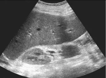
Marked, homogeneous hepatomegaly with uniform extension below the lower costal margin.
Diffuse increased parenchymal echogenicity ('bright liver') creating increased contrast with the retrohepatic structures.
:::note[Diagnostic Pearls]
- Type I (von Gierke): Often presents with massive hepatomegaly alongside concurrent nephromegaly, as the renal cortex also stores excess glycogen.
- Type III (Cori): Combines severe hepatomegaly with systemic skeletal muscle weakness.
- The underlying liver parenchyma frequently remains smooth and structurally uniform until secondary complications or late-stage cirrhosis develop. :::
Disease-Specific Findings
Different enzymatic deficiencies alter the long-term structural progression of the liver:
| GSD Type | Enzyme Deficiency | Key Ultrasound Features |
|---|---|---|
| Type I (von Gierke) | Glucose-6-phosphatase | Massive hepatomegaly, nephromegaly, multiple hepatic adenomas (frequently appearing after puberty) |
| Type III (Cori) | Glycogen debranching enzyme | Marked hepatomegaly; can progressively develop architectural cirrhosis during adulthood |
| Type IV | Branching enzyme | Progressive cirrhosis characterized by highly coarse, heterogeneous echotexture |
| Type VI / IX | Liver phosphorylase / kinase | Mild to moderate hepatomegaly that often spontaneously improves with patient age |
Hepatic Adenomas in GSD-I
Hepatic adenomas develop in 22 to 75 percent of GSD-I patients by their second or third decade of life:
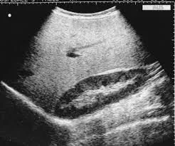
- They typically appear as well-circumscribed, hypoechoic focal masses against the bright background liver.
- Multiple separate lesions are common (hepatic adenomatosis).
- High-resolution surveillance imaging is strongly recommended every 6 to 12 months due to the documented risk of malignant transformation into hepatocellular carcinoma (HCC).
Monitoring and Complications
1. Adenoma Surveillance
- Establish a baseline abdominal ultrasound exam at puberty for all GSD-I patients.
- Strictly monitor for rapid size increases or lesions exceeding 5 cm, which carry a higher risk of spontaneous hemorrhage or malignant change.
- Utilize Contrast-Enhanced Ultrasound (CEUS), CT, or MRI if any focal mass displays atypical or suspicious features.
2. Progression to Cirrhosis
- Cirrhotic progression is most commonly seen in GSD types III and IV.
- Sonographers must carefully scan for a nodular capsular surface, highly coarse parenchymal remodeling, and secondary signs of portal hypertension (such as splenomegaly or ascites fluid collections).
3. Systematic Ultrasound Protocol for GSD
When performing a follow-up assessment on a GSD patient, always ensure you cover these structural steps:
- Measure and record the absolute vertical liver span in the midclavicular line.
- Formally grade the parenchymal echogenicity by comparing it directly to the right renal cortex.
- Screen all segments using high-frequency transducers to detect small, early adenomatous nodules.
- Assess the portal vein flow direction and diameter if signs of chronic architectural damage are present.
- Extend the survey to include absolute longitudinal measurements of both kidneys (essential for GSD-I).