Hydrocele: Ultrasound Diagnosis & Evaluation
Hydrocele refers to an abnormal accumulation of serous fluid between the parietal and visceral layers of the tunica vaginalis. The normal scrotum contains a small amount of fluid, which is often visible on ultrasound. However, larger fluid collections may develop due to various congenital or acquired causes.
These collections are typically confined to the anterolateral aspects of the scrotum because of the posterior attachment of the testis to the epididymis and scrotal wall (bare area).
Clinical Presentation
Hydrocele is the most common cause of painless scrotal swelling. It may be:
- Congenital (common in infants)
- Acquired (more common in adults)
Patients may present with:
- Painless scrotal enlargement
- Heaviness or discomfort in the scrotum
- Difficulty palpating the testis (in large hydroceles)
Types of Hydrocele
-
Congenital Hydrocele:
Results from incomplete closure of the processus vaginalis, allowing communication between the peritoneal cavity and scrotum. This often resolves spontaneously by 18 months of age. -
Acquired Hydrocele:
May be idiopathic or secondary to:- Epididymitis
- Epididymo-orchitis
- Testicular torsion
- Trauma
- Tumors (usually associated with small hydroceles)
-
Encysted (Noncommunicating) Hydrocele:
Loculated fluid collection along the spermatic cord without communication with the peritoneum. -
Funicular Hydrocele:
Communicates with the peritoneum but not with the scrotal sac.
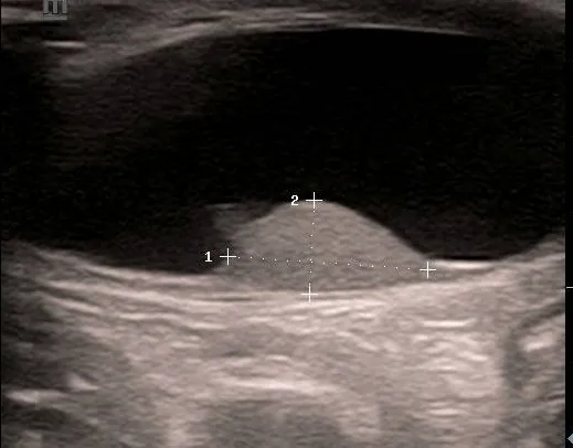
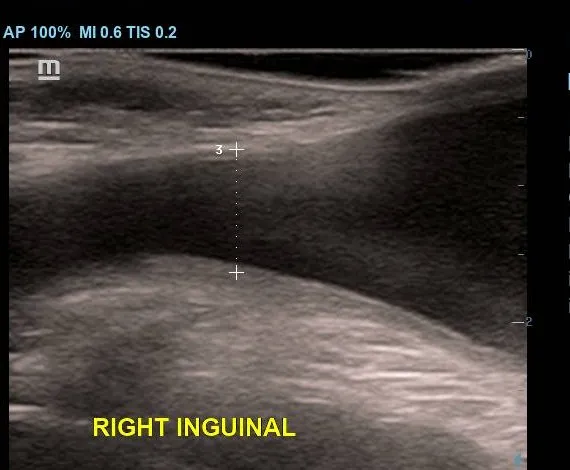
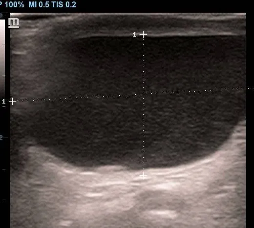
Ultrasound Features
-
Anechoic fluid collection: Surrounding the anterolateral aspects of the testis
-
Well-defined margins: Confined within the tunica vaginalis
-
Posterior acoustic enhancement: Due to fluid content
-
Internal echoes (occasionally):
- Low-level echoes from fibrin strands
- Cholesterol crystals moving freely within the fluid
-
Mass effect: Large hydroceles may obscure testicular evaluation clinically
-
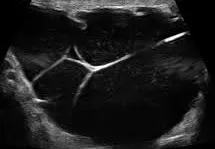
Rare Doppler finding:
Very large hydroceles may impair venous drainage, leading to reduced or absent diastolic arterial flow
:::note[Key Diagnostic Points]
- Anechoic fluid surrounding the testis is characteristic of a simple hydrocele
- Internal echoes suggest proteinaceous content or complications
- Always evaluate the underlying testis for associated pathology
:::
Complicated Fluid Collections
Hematocele
Hematocele refers to accumulation of blood within the tunica vaginalis and may result from:
- Trauma
- Surgery
- Testicular torsion
- Neoplasms
Ultrasound Features:
- Complex fluid with internal echoes
- Septations and clot formation
- May evolve over time with increasing echogenicity
Pyocele
Pyocele is a collection of pus within the tunica vaginalis, usually due to:
- Untreated epididymo-orchitis
- Ruptured intratesticular abscess
Ultrasound Features:
-
Complex heterogeneous fluid
-
Internal septations and debris
-
Possible gas echoes in severe infection
-
Associated findings:
- Thickened scrotal wall
- Hyperemia on Doppler
- Calcifications in chronic cases
Role of Ultrasound
Ultrasound plays a crucial role in:
- Confirming the diagnosis of hydrocele
- Differentiating simple from complex fluid collections
- Evaluating the testis when physical examination is limited
- Detecting underlying causes such as infection, torsion, or tumor
Early and accurate imaging helps guide appropriate management and prevents missed diagnoses of serious underlying conditions.