Peribiliary Cysts: Ultrasound Diagnosis & Management
Peribiliary cysts are rare, benign cystic lesions that occur in the hepatic hilum and along the larger intrahepatic bile ducts. These cysts develop from the peribiliary glands and are typically discovered incidentally during imaging studies for unrelated conditions.
While often asymptomatic, peribiliary cysts can sometimes lead to biliary obstruction or be mistaken for more serious conditions, making their recognition on ultrasound important for proper patient management.
Key Diagnostic Tips:
- Use a high-frequency linear probe for the best structural resolution.
- Always assess the spleen (up to 80 percent of diffuse fungal or systemic microlesions have concurrent splenic presence).
- Follow-up scans should preserve identical technical parameters for tracking accuracy.
Ultrasound Features
On ultrasound, peribiliary cysts appear as multiple small, round or oval anechoic structures clustered around the portal triads. They demonstrate through-transmission (posterior acoustic enhancement) and lack internal echoes or septations. The cysts are typically arranged linearly along the course of the bile ducts.
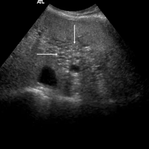
Key Ultrasound Characteristics
- Multiple small anechoic cysts (typically 0.2 to 2.5 cm in diameter)
- Strict localization along the portal triads (hepatic hilum and large intrahepatic bile ducts)
- No communication with the biliary tree (distinguishing them from Caroli disease)
- Thin, smooth walls devoid of internal septations or solid components
- Present posterior acoustic enhancement
- Total absence of internal vascularity on Doppler examination
Differential Diagnosis
| Condition | Distinguishing Features |
|---|---|
| Caroli disease | Communicates directly with the biliary tree; may show the characteristic “central dot” sign |
| Biliary hamartomas | Displays a more diffuse liver distribution; smaller size profile (usually less than 1 cm) |
| Simple hepatic cysts | Random parenchymal distribution; completely unassociated with portal triads |
| Periportal lymphadenopathy | Solid tissue appearance; frequently demonstrates internal vascularity on Doppler |
Associated Systemic Conditions: Peribiliary cysts are much more commonly identified in patients suffering from:
- Liver cirrhosis (particularly within advanced structural stages)
- Portal hypertension
- Polycystic liver disease
- Congenital hepatic fibrosis
Management Approach
1. Clinical Action Plans
- Asymptomatic cysts: No treatment needed; standard periodic monitoring may be safely considered.
- Symptomatic cysts: Consider interventional drainage or surgical evaluation if the lesions cause mechanical biliary obstruction.
- Uncertain diagnosis: Follow-up imaging or additional modalities (such as contrast CT or MRCP) are strongly warranted.
- Associated conditions: Focus primarily on managing the patient’s underlying liver disease.
2. Follow-up Recommendations
- Typical and Asymptomatic: No routine imaging follow-up required.
- Atypical or New Symptoms: Consider a targeted follow-up ultrasound in 6 to 12 months, or upgrade to advanced imaging modalities.
- Underlying Liver Disease: Follow regular staging intervals as indicated by the primary disease process.
3. Interventional Options
For severe, symptomatic cases causing confirmed biliary obstruction:
- Percutaneous cyst drainage (effective as a temporary stabilizing measure)
- Endoscopic cyst unroofing or marsupialization
- Surgical resection (rarely required; reserved explicitly for refractory cases)
References
- Nakanuma Y, Kurumaya H, Ohta G. Multiple cysts in the hepatic hilum and their pathogenesis. A suggestion of periductal gland origin. Virchows Arch A Pathol Anat Histopathol. 1984;404(4):341-350.
- Itai Y, Ebihara R, Tohno E, et al. Hepatic peribiliary cysts: multiple tiny cysts within the larger portal tract, hepatic hilum, or both. Radiology. 1994;191(1):107-110.
- Terada T, Nakanuma Y. Pathological observations of intrahepatic peribiliary glands in 1,000 consecutive autopsy livers. III. Survey of necroinflammation and cystic dilatation. Hepatology. 1990;12(5):1229-1233.
- Wachsberg RH. Peribiliary cysts: a benign cause of bile duct dilatation that may mimic obstruction. AJR Am J Roentgenol. 1997;168(2):501-502.
- Baron RL, Campbell WL, Dodd GD 3rd. Peribiliary cysts associated with severe liver disease: imaging-pathologic correlation. AJR Am J Roentgenol. 1994;162(3):631-636.