Polycystic Liver Disease: Ultrasound Diagnosis & Management
Polycystic liver disease (PLD) is a genetic disorder characterized by the presence of multiple cysts scattered throughout the liver parenchyma. While often associated with autosomal dominant polycystic kidney disease (ADPKD), PLD can also occur as an isolated condition (autosomal dominant polycystic liver disease, ADPLD). Ultrasound plays a crucial role in diagnosis, monitoring, and management of this condition.
Key Diagnostic Tips:
- Use a high-frequency linear probe for the best structural resolution.
- Always assess the spleen (up to 80 percent of patients can have concurrent splenic cystic lesions).
- Follow-up scans should preserve identical technical parameters for precise tracking accuracy.
Ultrasound Features
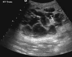
On ultrasound, early-stage PLD is characterized by clean, anechoic structures with distinct borders. As the condition advances, the healthy liver tissue becomes increasingly compressed and replaced by escalating numbers of expanding cysts.
Characteristic Ultrasound Features
- Multiple cysts: Typically greater than 20 cysts distributed throughout both liver lobes
- Cyst morphology: Round or oval anechoic structures with sharp margins
- Cyst size: Varies from a few millimeters to several centimeters
- Cyst walls: Thin, smooth walls (less than 1mm thickness)
- Acoustic enhancement: Pronounced posterior acoustic enhancement behind cysts
- No internal flow: Complete absence of internal vascularity on Doppler evaluation
- Parenchymal changes: Normal liver parenchyma between cysts in early stages
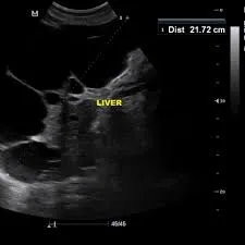
Advanced Disease Features
In advanced stages of polycystic liver disease, ultrasound evaluations typically demonstrate:
- Massive hepatomegaly resulting in secondary abdominal distension
- Near-complete replacement of functional liver parenchyma by clustered cysts
- Mechanical compression of adjacent macrostructures (Inferior Vena Cava, portal vein, and bile ducts)
- Complicated cyst profiles showcasing internal hemorrhage, infection, or frank wall rupture
- Secondary signs of portal hypertension in progressive, severe cases
Diagnostic Criteria
Ultrasound Diagnostic Criteria for PLD
For ADPKD-associated PLD:
- Age less than 40 years: 3 or more liver cysts
- Age 40 to 59 years: 4 or more liver cysts
- Age 60 years or older: 5 or more liver cysts
For isolated ADPLD:
- Age less than 40 years: 4 or more liver cysts
- Age 40 years or older: 10 or more liver cysts
- Total absence (or a very low, isolated profile) of renal cysts
- Documented family history of polycystic liver disease
Grading System for Disease Severity
| Grade | Description | Ultrasound Findings |
|---|---|---|
| I (Mild) | Less than 10 cysts | Normal liver architecture, all cysts remain less than 5cm |
| II (Moderate) | 10 to 20 cysts | Mild parenchymal replacement, select dominant cysts exceed 5cm |
| III (Severe) | Greater than 20 cysts | Significant parenchymal replacement (greater than 50% of liver volume) |
| IV (Massive) | Innumerable cysts | Near-total parenchymal replacement with marked gross hepatomegaly |
Differential Diagnosis
| Condition | Distinguishing Features | Key Differences from PLD |
|---|---|---|
| Simple hepatic cysts | Solitary or few isolated cysts | Significantly fewer in number; total lack of underlying genetic basis |
| Biliary hamartomas | Multiple small microcysts (less than 1.5cm) | Uniformly small size profile; completely lacks a family history |
| Caroli disease | Segmental cystic biliary dilatation | Communicates directly with the biliary tree; features the central dot sign |
| Hydatid cysts | Complex, layered cystic masses | Presence of internal daughter cysts and secondary wall calcifications |
| Metastatic disease | Complex or necrotic cystic masses | Thickened irregular walls, solid mural components, known primary malignancy |
Clinical Implications
Complications Detectable by Ultrasound
- Cyst hemorrhage: New internal low-level echoes and changing fluid-debris levels
- Cyst infection: Noticeably thickened walls, internal echoes, and occasional gas bubbles
- Biliary obstruction: Secondary dilatation of the upstream intrahepatic bile ducts
- Vascular compression: Narrowed or displaced hepatic veins, main portal vein, or Inferior Vena Cava
- Cyst rupture: Ill-defined cyst margins accompanied by free intraperitoneal fluid
Monitoring Recommendations
- Establish an initial baseline ultrasound for all newly diagnosed ADPKD patients.
- Schedule an annual ultrasound for symptomatic individuals or those categorized with Grade III/IV disease severity.
- Perform screening scans every 2 to 3 years for stable, asymptomatic Grade I/II disease presentations.
- Order an immediate targeted ultrasound if acute symptoms appear (such as localized pain, sudden fever, or jaundice).
- Utilize detailed ultrasound mappings before scheduling any planned surgical or percutaneous interventions.
References
- Gevers TJ, Drenth JP. Diagnosis and management of polycystic liver disease. Nat Rev Gastroenterol Hepatol. 2013;10(2):101-108.
- Van Aerts RMM, Van de Laarschot LFM, Banales JM, Drenth JPH. Clinical management of polycystic liver disease. J Hepatol. 2018;68(4):827-837.
- Hoevenaren IA, Wester R, Schrier RW, et al. Polycystic liver: clinical characteristics of patients with isolated polycystic liver disease compared with patients with polycystic liver and autosomal dominant polycystic kidney disease. Liver Int. 2008;28(2):264-270.
- Qian Q, Li A, King BF, et al. Clinical profile of autosomal dominant polycystic liver disease. Hepatology. 2003;37(1):164-171.
- Mavilia MG, Pakala T, Molina M, Wu GY. Differentiating Cystic Liver Lesions: A Review of Imaging Modalities, Diagnosis and Management. J Clin Transl Hepatol. 2018;6(2):208-216.
- European Association for the Study of the Liver. EASL Clinical Practice Guidelines on the management of cystic liver diseases. J Hepatol. 2022;77(4):1083-1108.