Prostate Ultrasound
Prostate ultrasound is an essential imaging modality for evaluating prostate pathology due to detailed visualization of zonal architecture, real-time guidance for interventions, the absence of ionizing radiation, and the ability to assess vascularity with Doppler. However, it remains operator-dependent and has limited extracapsular evaluation compared to MRI.
Prostate Anatomy
Zonal Anatomy
- Peripheral zone: 70% of glandular tissue, hypoechoic, site of most cancers.
- Central zone: 25% of gland, hyperechoic, rarely involved by cancer.
- Transition zone: 5% (in young men), site of BPH, mixed echogenicity.
- Anterior fibromuscular stroma: Hypoechoic, non-glandular.
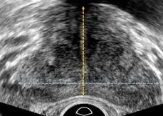
Normal Measurements
- Volume: 20-30 mL (calculated as $L \times W \times H \times 0.52$).
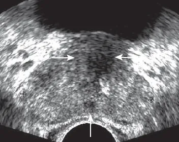
Transverse view at midgland level showing normal muscular internal urethral sphincter and ejaculatory ducts.
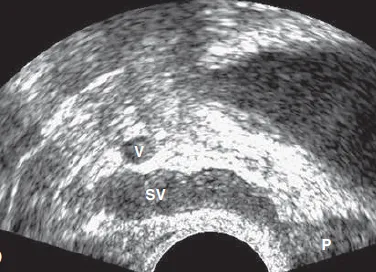
Sagittal view showing hypoechoic seminal vesicles superior to the prostate.
Clinical Indications
- Common Indications: Evaluation of prostate size in BPH, suspected prostate cancer, elevated PSA, prostatitis assessment, and guidance for biopsy[cite: 1].
- Clinical Scenarios: Includes management of LUTS/urinary retention, abnormal DRE, pelvic pain, and infertility evaluation.
Scanning Technique
- Patient Preparation: Bowel prep and antibiotic prophylaxis for TRUS; full bladder for transabdominal scans.
- Approach: Systematic survey in sagittal and transverse sweeps, utilizing 12-core sampling for biopsies.
Pathological Findings
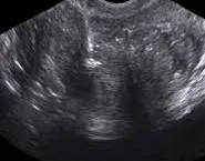
BPH showing enlarged transition zone and nodular appearance.
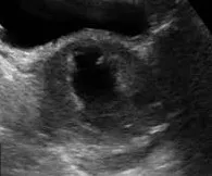
Prostate cancer presenting as a hypoechoic nodule in the peripheral zone.
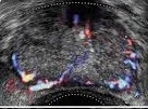
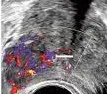
Acute prostatitis showing diffuse hypoechogenicity and increased vascularity.
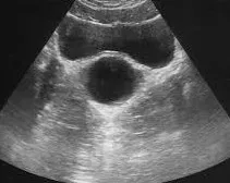
Well-defined anechoic cyst with posterior acoustic enhancement.
Echogenic foci with shadowing common in chronic prostatitis.
Hypoechoic hematoma following biopsy.
References
- American College of Radiology (ACR). (2023). ACR Appropriateness Criteria® Prostate Cancer Detection. Journal of the American College of Radiology, 20(1S), S78-S92.[cite: 1]
- Sidhu, P. S., et al. (2023). Clinical Ultrasound (4th ed.). Elsevier.
- European Society of Urogenital Radiology (ESUR). (2022). Guidelines on Prostate Ultrasound. European Radiology, 33(3), 261-279.
- Rifkin, M. D. (2023). Prostate Ultrasound. In: Rumack, C. M., & Levine, D. (Eds.), Diagnostic Ultrasound (6th ed., pp. 112-128). Elsevier.
- African Society of Uroradiology (ASUR). (2023). Consensus Guidelines on Prostate Ultrasound in African Populations. African Journal of Radiology, 28(1), 45-60.