Scrotal Ultrasound
Diagnostic ultrasound is the most common imaging technique used to supplement the physical examination of the scrotum and is an accurate means of evaluating many scrotal processes[cite: 1]. Technical advancements in high-resolution real-time sonography and color Doppler have expanded clinical applications to include the assessment of scrotal masses, acute pain, trauma, infertility, tumors, and undescended testis[cite: 1].
Scrotal Anatomy
Normal Structures
- Testis: Homogeneous medium-level echogenicity, with a length of 3-5 cm.
- Epididymis: Head (10-12 mm), body, and tail; iso- or hypoechoic to the testis.
- Tunica albuginea: Hyperechoic outer covering.
- Mediastinum testis: Seen as an echogenic band in the sagittal plane.
Normal Measurements
- Testis volume: 15-25 mL (calculated as $L \times W \times H \times 0.52$).
- Epididymal head: $\le 12$ mm.
- Pampiniform plexus veins: $\le 2$ mm diameter.
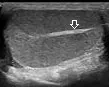
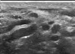
Longitudinal view showing homogeneous echotexture with mediastinum testis.
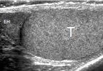
Longitudinal view showing epididymal head with slightly coarser echotexture compared to the testis.
Clinical Indications
- Common Indications: Acute scrotal pain (torsion, epididymitis), palpable scrotal mass, testicular trauma, infertility evaluation, and undescended testis localization[cite: 1].
- Specific Scenarios: Includes evaluation of testicular torsion, epididymo-orchitis, testicular cancer, varicocele, and hydrocele[cite: 1].
Scanning Technique
- Preparation: No specific preparation is required; however, a warm gel and scrotal support improve patient comfort.
- Equipment: Use a high-frequency linear transducer (7-15 MHz) with harmonic imaging enabled.
- Approach: Perform a systematic survey of both testes in two planes, comparing echotexture and vascularity bilaterally, and document any abnormalities.
Pathological Findings
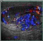
Testicular torsion showing absent intratesticular flow and whirlpool sign.
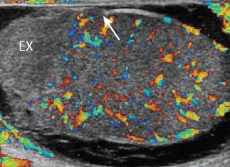
Enlarged hyperemic epididymis and increased testicular vascularity.
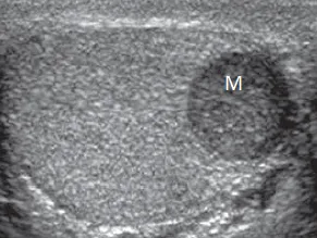
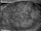
Hypoechoic intratesticular mass with increased vascularity.
Dilated pampiniform veins with increased flow on Valsalva.
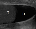
Anechoic peritesticular fluid without internal vascularity.
Disrupted tunica albuginea with heterogeneous echotexture.
References
- American College of Radiology (ACR). (2023). ACR Appropriateness Criteria® Acute Onset of Scrotal Pain. Journal of the American College of Radiology, 20(1S), S112-S125.
- Sidhu, P. S., et al. (2023). Clinical Ultrasound (4th ed.). Elsevier.
- European Society of Urogenital Radiology (ESUR). (2022). Guidelines on Scrotal Ultrasound. European Radiology, 33(3), 280-295.
- Dogra, V. S. (2023). Scrotal Ultrasound. In: Rumack, C. M., & Levine, D. (Eds.), Diagnostic Ultrasound (6th ed., pp. 129-145). Elsevier.
- African Society of Uroradiology (ASUR). (2023). Consensus Guidelines on Scrotal Ultrasound in African Populations. African Journal of Radiology, 28(1), 61-75.