Neck & Thyroid Ultrasound
Neck ultrasound is the primary imaging modality for evaluating thyroid and parathyroid pathology, as well as cervical lymph nodes and other neck structures. High-resolution ultrasound provides detailed assessment of thyroid nodules, inflammatory conditions, and malignancies. Doppler evaluation adds functional information about vascular patterns that helps differentiate benign from malignant lesions. In Africa, where thyroid disorders are prevalent due to both iodine deficiency and autoimmune conditions, ultrasound plays a crucial role in diagnosis and management.
Key advantages
- No ionizing radiation exposure
- Real-time imaging with high spatial resolution
- Ability to guide fine needle aspiration biopsies
- Assessment of vascular patterns with Doppler
- Evaluation of adjacent structures (lymph nodes, parathyroids)
Neck Anatomy
Normal Thyroid
- Homogeneous medium-level echogenicity (slightly hyperechoic to strap muscles)
- Butterfly shape with isthmus connecting two lobes
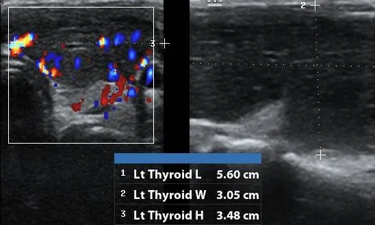
- Normal measurements: Lobe length 4-6cm, AP 1.5-2cm, width 2-2.5cm
- Isthmus thickness: ≤5mm
- Normal vascularity on Doppler (mainly inferior thyroid arteries)
Adjacent Structures
- Trachea: Hyperechoic anterior wall with posterior shadowing
- Common carotid artery: Lateral to thyroid lobe
- Internal jugular vein: Lateral to carotid, compressible
- Parathyroid glands: Normally not visible, may be seen when enlarged
- Cervical lymph nodes: Oval shape with fatty hilum when normal
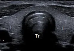
Transverse view showing homogeneous thyroid lobes (T) with trachea (Tr) and common carotid artery (CCA).
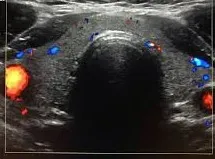
Color Doppler showing normal parenchymal flow in thyroid lobes.
Clinical Indications
-
Common Indications
- Evaluation of palpable neck mass
- Assessment of thyroid nodules
- Follow-up of known thyroid disease
- Guidance for fine needle aspiration biopsy
- Evaluation of suspected thyroiditis
-
Specific Clinical Scenarios
- Thyroid nodules: Risk stratification using TI-RADS
- Thyroid cancer: Evaluation of primary tumor and lymph nodes
- Graves’ disease: Hypervascular “thyroid inferno” pattern
- Hashimoto’s thyroiditis: Heterogeneous, hypoechoic parenchyma
- Parathyroid adenoma: Hypoechoic oval lesion posterior to thyroid
Scanning Technique
- Patient Prep: Supine with neck extended, remove neck jewelry.
- Equipment: High-frequency linear transducer (7-15 MHz), harmonic imaging enabled.
- Approach: Systematic survey of both lobes in transverse and longitudinal planes. Measure isthmus, assess nodules, and evaluate cervical lymph nodes (levels II-VI).
Pathological Findings
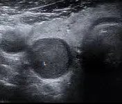
Benign nodule showing cystic components and spongiform appearance.

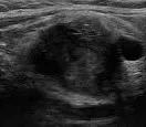
Malignant nodule: Hypoechoic, microcalcifications, taller-than-wide.
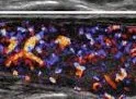
Graves' disease showing enlarged gland and hypervascular 'thyroid inferno'.
Hashimoto's thyroiditis: Heterogeneous echotexture and hypoechoic parenchyma.
Parathyroid adenoma appearing as a hypoechoic rounded lesion posterior to the thyroid.
Metastatic lymph node: Round shape with loss of fatty hilum and cystic changes.
References
- American College of Radiology (ACR). (2023). ACR TI-RADS: Thyroid Imaging, Reporting and Data System. Journal of the American College of Radiology.
- Tessler, F. N., et al. (2023). Thyroid and Parathyroid Ultrasound (2nd ed.).
- European Thyroid Association (ETA). (2022). Guidelines on Thyroid Ultrasound. European Thyroid Journal.
- African Society of Endocrine Surgeons (ASES). (2023). Consensus Guidelines on Thyroid Ultrasound in African Populations. African Journal of Endocrine Surgery.