Uterus Ultrasound
Uterine ultrasound is the primary imaging modality for evaluating gynecological pathology due to its excellent visualization of anatomy, real-time assessment of the endometrium, lack of ionizing radiation, and cost-effectiveness in resource-limited settings[cite: 1]. While powerful, it remains operator-dependent and has limitations in evaluating deep myometrial invasion compared to MRI[cite: 1].
Uterine Anatomy
Layers of the Uterus
- Endometrium: The inner mucosal layer; its echogenicity varies with the menstrual cycle.
- Myometrium: The middle muscular layer, exhibiting homogeneous mid-level echogenicity.
- Serosa: The outer layer, visualized as a thin hyperechoic line.
Normal Measurements
- Nulliparous: 6-8 cm length, 3-5 cm width, 2-3 cm AP.
- Multiparous: 8-10 cm length, 4-6 cm width, 3-5 cm AP.
- Postmenopausal: $\le 6$ cm length, $\le 2$ cm AP (without HRT).
- Endometrial thickness: Varies by cycle phase (4-16 mm) and is $\le 4$ mm in postmenopausal patients.
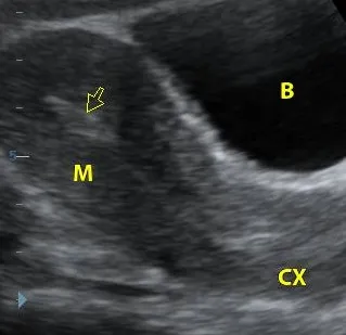
Midline sagittal view showing normal endometrial stripe, myometrium, and cervix.

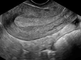
Proliferative phase endometrium with a triple-layer appearance.
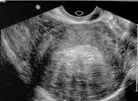
Secretory phase endometrium with thick, uniform hyperechoic appearance.
Clinical Indications
- Common Indications: Abnormal uterine bleeding, pelvic pain, suspected fibroids or adenomyosis, infertility, and postmenopausal bleeding[cite: 1].
- Clinical Scenarios: Evaluation of bulk symptoms from fibroids, dysmenorrhea in adenomyosis, endometrial cancer screening, and congenital anomalies[cite: 1].
Scanning Technique
- Preparation: Full bladder for transabdominal scans; empty bladder for transvaginal scans.
- Approach: Perform systematic sagittal and transverse sweeps. The optimal time for endometrial assessment is during days 5-10 of the menstrual cycle.
- Equipment: Use curvilinear transducers (3-5 MHz) for transabdominal imaging and endovaginal transducers (5-9 MHz) for TVS.
Pathological Findings
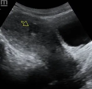
Well-defined hypoechoic fibroid with posterior acoustic shadowing.
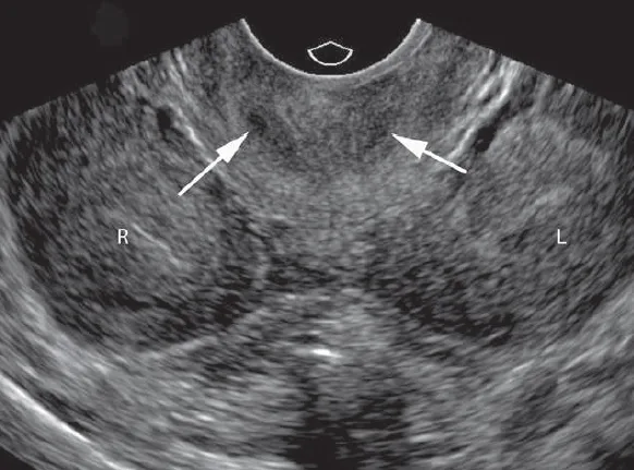
Diffuse uterine enlargement with heterogeneous myometrium and cysts.
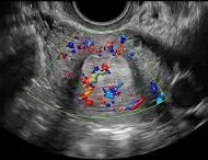
Focal endometrial thickening with a feeding vessel (pedicle sign).
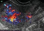
Thickened, irregular endometrium with increased chaotic vascularity.
Uterus didelphys showing two separate cavities and two cervices.
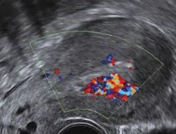
Heterogeneous endometrial mass with vascularity post-delivery.
References
- American College of Radiology (ACR). (2023). ACR Appropriateness Criteria® Abnormal Uterine Bleeding. Journal of the American College of Radiology, 20(1S), S78-S92.
- Salen, P., et al. (2023). Gynecologic Ultrasound (4th ed.). Elsevier.
- European Society of Urogenital Radiology (ESUR). (2022). Guidelines on Gynecological Ultrasound. European Radiology, 33(3), 261-279.
- Timor-Tritsch, I. E. (2023). Uterine Ultrasound. In: Rumack, C. M., & Levine, D. (Eds.), Diagnostic Ultrasound (6th ed., pp. 112-128). Elsevier.
- African Society of Uroradiology (ASUR). (2023). Consensus Guidelines on Gynecological Ultrasound in African Populations. African Journal of Radiology, 28(1), 45-60.